Axial length changes from retinal detachment surgery. Age 52.
| Sphere | Cylinder | Axis | Add | PD | |
| OD | -1.50 | 2.00 | 32 | ||
| OS | +4.00 | 2.00 | 32 |
| BU | BD | BI | BO | ||
| 2.5 | 2.5 | 4 | 12 |
| Eye | DBL | Wrap | Vertex | Height | |
| 56 | 16 | 6 | 12 | 22 |
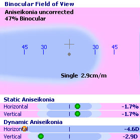
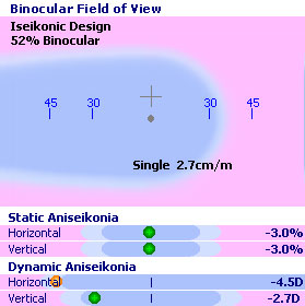
This patient had a sudden refractive error change subsequent to retinal detachment surgery with a scleral buckle. It is common for axial length changes due to the procedure to create myopic anisometropia. The optometrist was assisted by the SHAW lens design tool, which enabled the clinician to easily specify the iseikonic correction. This method optimizes the design to accommodate the unique motor fusion physiology of each patient without resorting to bicentric manufacturing.


The -3% the doctor specifies for static magnification is achieved without a detrimental effect on dynamic aniseikonia. Note how the area of single vision compares to a conventional design while sensory fusion is maintained. This is accomplished by base curve optimization with minimal thickness penalty (max ET 4.2 mm). Vertex changes with base curvature and the non-linearity of base effects contribute to a design that is not predictable using paraxial methods.